POTASSIUM HYDROXIDE (KOH) WET MOUNT
AIM
To visualize the fungal element
present in the Clinical specimens (Skin, Hair and Nail).
PRINCIPLE
Potassium hydroxide (KOH)
preparation is used for the rapid detection of fungal elements in a clinical
specimens, as it clears the specimen making fungal elements more visible during
Direct microscopic examination. Potassium hydroxide (KOH) solution is
alkaline and has the ability to dissolve keratin that is scraped from the outer
layer of the skin or nail or hair. As the KOH dissolves, the material binding
the skin cells together, any fungus present is released. This allows for the
identification of organisms such as Dermatophytes.
The
affected Skin or Nail is gently scraped with a small Scalpel or the edge of a
Glass slide. The scrapings from the skin are placed on a microscope slide and a
few drops of a Potassium hydroxide (10 – 20 % KOH) solution are added. A
coverslip is placed over the KOH-digested sample and the slide is heated for a
short time. The slide was incubated for 5 – 10 minutes. The slide is examined under
the microscopically without staining. Potassium hydroxide is a highly
corrosive deliquescent chemical; therefore it should be handled
with great care.
MATERIALS REQUIRED
- Clinical specimens (Skin, Nail, Hair or Sputum)
- Potassium hydroxide (KOH)
- Scalpel
- Bunsen burner
- Teasing needle
- Glass slide
- Coverslip
- Microscope
PROCEDURE
a) Place the Clinical specimen (Skin, Nail, Hair or
Sputum) on the clean slide.
b) Pour a drop of 10 – 20 % KOH on the specimen and place
a coverslip over it.
c) Heat the KOH digested sample containing slide gently
over the flame.
d) Incubate the slide for 5 - 10 minutes or place the
slide in a petridish, or other containers with a lid, together with a damp
piece of filter paper or cotton wool to prevent the preparation from drying
out.
e) Observe the slide without any staining under
microscope at 40x objective lens.
OBSERVATION AND RESULTS
- Different fungal elements like hyphae, pseudohyphae, yeast cells, spores, spherules, and sclerotic bodies can be seen clearly in a KOH wet mount.
- In dermatophytosis, arthrospores develop and form as hyphae break apart and appear as a linear chain of small, round to rectangular, highly refractile structures.
- In KOH preparations of sputum, the fungus appears as non-pigmented septate hyphae, 3 – 5 µm in diameter, with characteristic dichotomous branching and an irregular outline.
- KOH preparation is used for the diagnosis of ringworm infection. Laboratory diagnosis of Tinea rests on the identification of an organism by microscopic examination of skin or nail scrapings with 10 % to 20 % KOH on wet mount examination.
- KOH with blue-black ink preparation is recommended if Malassezia furfur is suspected.
- Calcofluor White–Potassium Hydroxide Preparation can also be used for the examination of Fungal infection because Calcofluor White is a non-specific stain, an appreciation for fungal element morphology on direct examination is crucial for adequate specimen interpretation.
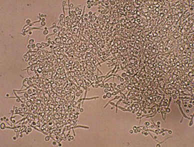
Figure – 1: KOH wet mount of Candida albicans
Comments
Post a Comment