
LACTOPHENOL COTTON BLUE (LPCB) STAINING

 LACTOPHENOL COTTON BLUE (LPCB) STAINING


AIM

           To identify the Microscopic morphology of Fungi by Lactophenol Cotton Blue (LPCB) Staining.

PRINCIPLE  

            Fungi are the Eukaryotic microorganisms which are classified into two main group viz., (i) Molds (Multicellular) and (ii) Yeasts (Unicellular). The Lactophenol Cotton Blue (LPCB) is a stain used for making semi-permanent microscopic preparation of Fungi. The LPCB stain has three components: (i) Phenol – Act as a Disinfectant or Fungicide and kills any living microorganisms; (ii) Lactic acid – Preserves the Fungal structure and (iii) Cotton blue stain - Stains the chitin and cellulose of the fungal cell wall intensely blue. LPCB solution is a mounting medium and staining agent used in the preparation of slides for microscopic examination of fungi. In LPCB staining, fungal elements are stained intensely in Blue colour.

MATERIALS REQUIRE

  • Fungal culture plate
  • Lactophenol Cotton Blue (LPCB) Stain
  • Bunsen burner
  • Teasing needle
  • Glass slide
  • Coverslip
  • Microscope.

PROCEDURE

  • Place a drop of Lactophenol Cotton Blue reagent on a clean, grease free and dry slide. The stain imparts a blue colouration on hyphae.
  • By using a Teasing needle, carefully tease the small portion of fungal culture into a thin preparation and form the Fungal mycelial mat.
  • Place a coverslip gently on the Fungal mycelial mat without air bubbles and wait for about 3 - 5 minutes.
  • Observe under microscope at 40 X objective lens for detailed examination of Fungal morphology and Spores.

OBSERVATION AND RESULTS

        The morphology of the fungi appears (mycelium and spores) as dark blue stained mycelium under microscope in 45 X objective.


Figure – 1: Microscopic morphology of Aspergillus niger


Figure –  2: Microscopic morphology of Aspergillus flavus


Figure – 3: Microscopic morphology of Aspergillus fumigatus


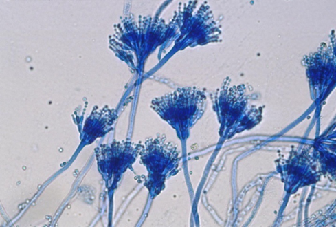
Figure – 4: Microscopic morphology of Penicillium chrysogenum

Figure – 5: Microscopic morphology of Rhizopus sp. (Rhizoids present) 

Figure – 6: Microscopic morphology of Mucor sp. (Rhizoids absent)

Comments

Popular posts from this blog

International Online Short Term Certificate Course on “SPECTROSCOPY – AN OVERVIEW” (Phase - I) (for Faculties, Research Scholars and Students)

  DAY CHAPTER NUMBER TOPICS Day – 1 06.01.2025 1 An Introduction to Spectroscopy 2 History of Spectroscopy 3 Types of Spectroscopy 4 Spectroscopy – Applications, Advantages and Limitations Day – 2 07.01.2025 5 An Introduction to Spectrophotometer 6 Preventive Measures in Spectrophotometer 7 Spectrophotometer – Applications, Advantages & Limitations Day – 3 08.01.2025 8 Single Beam Spectrophotometer 9 Double Beam Spectrophotometer 10 Difference between Single Beam Spectrophotometer and Double Beam Spectrophotometer 11 Absorption Spectroscopy Day – 4 09.01.2025 12 Ultraviolet (UV) Spectroscopy 13 Ultravio...

15 Days International Online Certificate Course on “MICROBIAL INSTRUMENTATION – AN OVERVIEW” (Phase - I)

  DAY CHAPTER NUMBER TOPICS Day – 1 27.01.2025 1 Orientation to the Microbiology Laboratory 2 Parts of Microscope 3 Bright Field Microscope and Dark Field Microscope 4 Difference between Simple Microscope and Compound Microscope Day – 2 28.01.2025 5 Electron Microscope 6 Difference between Light Microscope and Electron Microscope 7 Difference between Scanning Electron Microscope and Transmission Electron Microscope Day – 3 29.01.2025 Quiz 1 from Day 1 Lecture Videos and Day 2 Lecture Videos (20 Multiple Choice Questions) Day – 4 30.01.2025 8 An Introduction to Spectrophotometer 9 Preventive Measures in Spectrophotometer 10 Spectrophotometer - Applications, Adva...

International Four Weeks Online Certificate Course on “MICROBIOLOGY LABORATORY TECHNIQUES” (Phase - III)

ONLINE COURSE CONTENTS DAY CHAPTER NUMBER TOPICS Week - 1 1 Orientation to Microbiology Laboratory 2 Sterilization – Physical method (Drying, Dry heat and Moist heat) 3 Sterilization – Physical method (Filtration and Radiation) 4 Sterilization – Chemical method 5 Evaluation of Disinfectants 6 Parts of Microscope 7 Difference between Simple microscope & Compound microscope 8 Difference between Light microscope & Electron microscope 9 Difference between SEM and TEM 10 Culture medium 11 Guidelines for the Collection of Clinical Specimens 12 Collection of Clinical specimens 13 Transport of Clinical specimens Week – 2 14 ...